
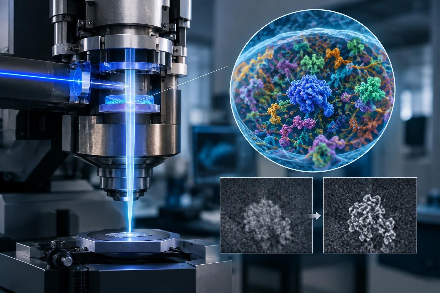
A cell is not a neat collection of parts laid out on a table. It is a crowded, shifting environment in which proteins and other molecules work amid countless neighbours. Seeing one small structure in that molecular bustle can be, as one researcher puts it, like trying to pick out a leaf in a forest.
A new microscope developed by physicists at the University of California, Berkeley, could help bring some of that hidden activity into view. Its key component, a laser phase plate, is designed to sharpen contrast in cryogenic electron microscopy, or cryo-EM. The goal is not simply to magnify more, but to make faint structures easier to distinguish from their surroundings.
Making subtle signals stand out
In conventional light microscopy, a longstanding problem is that many cell structures are nearly transparent. Because they absorb or scatter little light, they can be hard to see without staining—a process that kills the cells. In the 1930s, physicist Frits Zernike found a way to turn subtle changes in the phase of light waves into visible differences in brightness. His phase-contrast technique transformed cell imaging and earned him the 1953 Nobel Prize in Physics.
The Berkeley team has adapted that basic idea for an electron microscope. Instead of light passing through a specimen, cryo-EM uses electrons to reveal structures at much smaller scales, including proteins. The laser does not illuminate the sample. It interacts with the microscope’s electron beam, shifting its phase so that weak signals from small structures can become easier to detect.
That matters because many human proteins are too small to image clearly with current methods. Cryo-EM has helped researchers determine the shapes of biological molecules, knowledge that can support studies of disease and drug development. But a powerful microscope can still struggle when the signal from a tiny protein is lost against background noise. The researchers say their laser phase plate could make proteins as small as one-third the size of those that challenge current microscopes easier to image.
From isolated molecules to cellular neighbourhoods
The potential reach goes beyond producing sharper images of individual proteins. Cryo-electron tomography, or cryo-ET, combines views taken from different angles to build three-dimensional pictures of biological material in its cellular setting. That context is crucial: a molecule’s neighbours and position can help reveal how it works, not just what shape it has in isolation.
For cryo-ET, contrast is especially important. A cell interior is densely packed, and researchers must distinguish structures that may be small and close together. Better visibility could help scientists investigate how molecular components are arranged and interact in that environment. That is a possibility the technology may enable, not a discovery it has already made.
The first instrument built around the approach, named Theia, combines the laser phase plate with additional electron optics. A related microscope being developed at Biohub in Redwood City uses two lasers set at right angles, each operating at roughly half the power of the single-laser arrangement. The design is intended to reduce optical distortions and protect components from damage. Both teams are working with microscope manufacturer Thermo Fisher Scientific.
A promising tool, not a finished answer
The Berkeley team’s latest images and technical findings appear in the journal Science; details of the two-laser design have been shared in a preprint. The researchers describe the contrast improvement as a way to address a central limitation of cryo-EM and cryo-ET. The next test is how well the instruments perform across the varied, complicated samples scientists want to study.
If the approach proves useful in practice, it could give researchers a clearer view of proteins not as isolated shapes, but as participants in the crowded workings of a cell. That would not make the cell’s machinery simple. It would make more of its moving parts visible—and sometimes, seeing where a single leaf sits in the forest is the first step toward understanding the whole ecosystem.